

Beyond Pregnancy: Unveiling the Surprising Applications of Ultrasound Technology
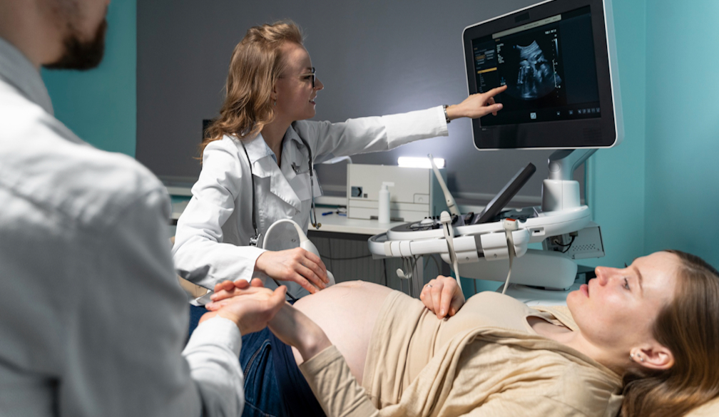
In the field of medical diagnostics, ultrasound technology has long been associated with monitoring pregnancies, offering expectant parents a glimpse into the development of their unborn child, and it is also crucial for procedures like ultrasound-guided biopsy. However, the utility of ultrasound extends far beyond obstetrics, delving into lesser-known areas of medical imaging. This article will help uncover the surprising applications of ultrasound technology, from musculoskeletal imaging to thyroid evaluation and vascular assessments.
Musculoskeletal Imaging
When you think of ultrasound, images of prenatal scans may come to mind. Yet, its capabilities extend to visualising the musculoskeletal system with remarkable clarity. This non-invasive technique allows healthcare professionals to assess soft tissue structures, joints, tendons, and ligaments. Whether diagnosing sports injuries, evaluating arthritis, or guiding therapeutic injections, this imaging method provides invaluable insights into the inner workings of the body's support system.
Thyroid Evaluation
The role of the thyroid gland is critical in regulating metabolism and hormone production. Disorders affecting the thyroid can have profound effects on health. Ultrasound imaging offers a non-radioactive and precise method for evaluating the thyroid gland and detecting abnormalities such as nodules, cysts, or enlargement. By visualising these structures in real-time, healthcare providers can diagnose conditions such as thyroid cancer, hyperthyroidism, or hypothyroidism early, facilitating timely intervention and treatment.
Breast Imaging
When screening for breast cancer, ultrasound is a valuable addition to mammography, especially for women who have dense breast tissue or are at a higher risk of developing the disease. It helps detect and characterise breast lesions, guiding further diagnostic evaluation and biopsy if necessary. It also aids in assessing the response to breast cancer treatment and monitoring for recurrence.
Vascular Assessments
Ultrasound technology transcends the sphere of static imaging, venturing into the dynamic domain of vascular assessments. By employing Doppler ultrasound, which measures the direction and speed of blood flow, doctors can evaluate the integrity of blood vessels. From detecting arterial blockages and assessing blood flow in the carotid arteries to monitoring varicose veins and identifying deep vein thrombosis, this imaging method serves as a versatile tool for diagnosing vascular conditions and guiding interventions.
Pelvic Floor Ultrasound
Pelvic floor ultrasound aids in diagnosing and assessing pelvic organ prolapse (POP) and urinary incontinence, common conditions affecting women. Visualising pelvic anatomy and measuring pelvic floor muscle function helps guide treatment decisions and monitor the effectiveness of interventions such as pelvic floor exercises or surgical repair.
Interventional Ultrasound
Ultrasound guidance enhances the safety and accuracy of various minimally invasive procedures, including biopsies, injections, and aspirations. By providing real-time imaging, it helps healthcare providers precisely target the desired area, reducing the risk of complications and improving procedural outcomes. This type of imaging is used in specialities such as radiology, oncology, pain management, and musculoskeletal medicine.
Accessibility and Versatility
What sets this imaging method apart from other imaging modalities is its accessibility and versatility. Unlike CT scans or MRIs, it does not expose patients to ionising radiation. This makes it safer for frequent use and ideal for certain populations, such as pregnant women and children. Ultrasound equipment is also portable, allowing for point-of-care imaging in diverse clinical settings, from rural clinics to emergency departments. If you want to get a deeper understanding of this subject, you can get mor information from Inside Radiology or from other reputed medical sites about the various applications of ultrasound.
Challenges and Future Directions
While ultrasound technology continues to revolutionise medical diagnostics, it is not without limitations. Factors such as operator dependency, limited penetration in obese patients, and difficulty visualising structures obscured by gas or bone can pose challenges. Nevertheless, ongoing advancements in technology, such as 3D and 4D imaging, contrast-enhanced ultrasound, and elastography, promise to overcome these obstacles, enhancing the diagnostic capabilities of this imaging method and expanding its applications even further.
The applications of ultrasound technology extend far beyond pregnancy. Using the power of sound waves, healthcare professionals can peer beneath the surface, uncovering a wealth of diagnostic information with precision and clarity. As people push the boundaries of medical imaging, embrace ultrasound technology to the fullest extent possible and realise its potential to revolutionise healthcare delivery and enhance patient outcomes.


