

Neuroscience in pictures: the best images of the year
- Written by: Wei Luan, Postdoctoral Researcher, The University of Queensland
To understand how the healthy brain works and what occurs in brain disease, neuroscientists use many microscopy techniques, ranging from whole-brain human MRIs to imaging within a single neuron (brain cell), creating stunning images in the process.
Here are a selection of the best and brightest produced by scientists at the Queensland Brain Institute at The University of Queensland in 2017.
 Wei 'Leon' Luan/QBI
This is a side view of a mouse embryo’s brain. The axons of neurons (dark blue) that release dopamine, a neurotransmitter involved in reward and pleasure, grow towards their target brain regions.
Wei 'Leon' Luan/QBI
This is a side view of a mouse embryo’s brain. The axons of neurons (dark blue) that release dopamine, a neurotransmitter involved in reward and pleasure, grow towards their target brain regions.
 Chase Sherwell/QBI
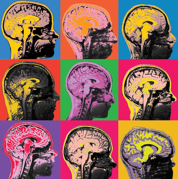
As neuroscience becomes increasingly of public interest, researchers are striving to make their findings accessible, with parallels to the pop art movement. These are MRI images of the human brain.
Chase Sherwell/QBI
As neuroscience becomes increasingly of public interest, researchers are striving to make their findings accessible, with parallels to the pop art movement. These are MRI images of the human brain.
 Abdalla Mohamed, PhD student/QBI
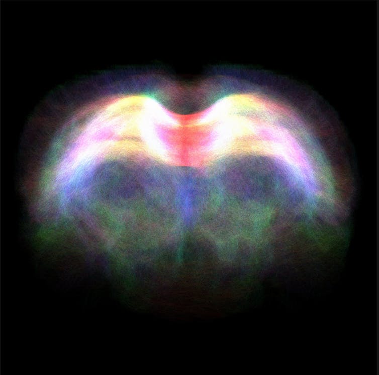
This image shows diffusion tensor imaging, an MRI-based neuroimaging technique, revealling the fibre tracts through the corpus callosum in a rodent brain. The corpus callosum links the brain’s left and right hemispheres to each other. The colours represent the different directions that the tracts are travelling through the brain.
Small-scale wonders
The colourful image below shows the nanoscale movements of individual molecules that are critical in mediating communication between neurons. Knowing how these molecules are organised, and how they move, is at the heart of understanding the brain in health and disease.
Abdalla Mohamed, PhD student/QBI
This image shows diffusion tensor imaging, an MRI-based neuroimaging technique, revealling the fibre tracts through the corpus callosum in a rodent brain. The corpus callosum links the brain’s left and right hemispheres to each other. The colours represent the different directions that the tracts are travelling through the brain.
Small-scale wonders
The colourful image below shows the nanoscale movements of individual molecules that are critical in mediating communication between neurons. Knowing how these molecules are organised, and how they move, is at the heart of understanding the brain in health and disease.
 Ravikiran Kasula/QBI
 Merja Joensuu/QBI
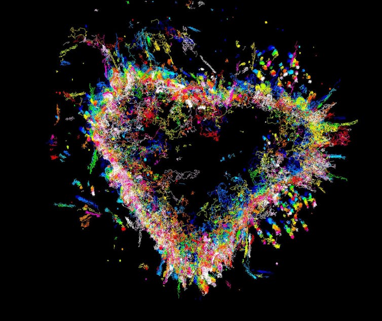
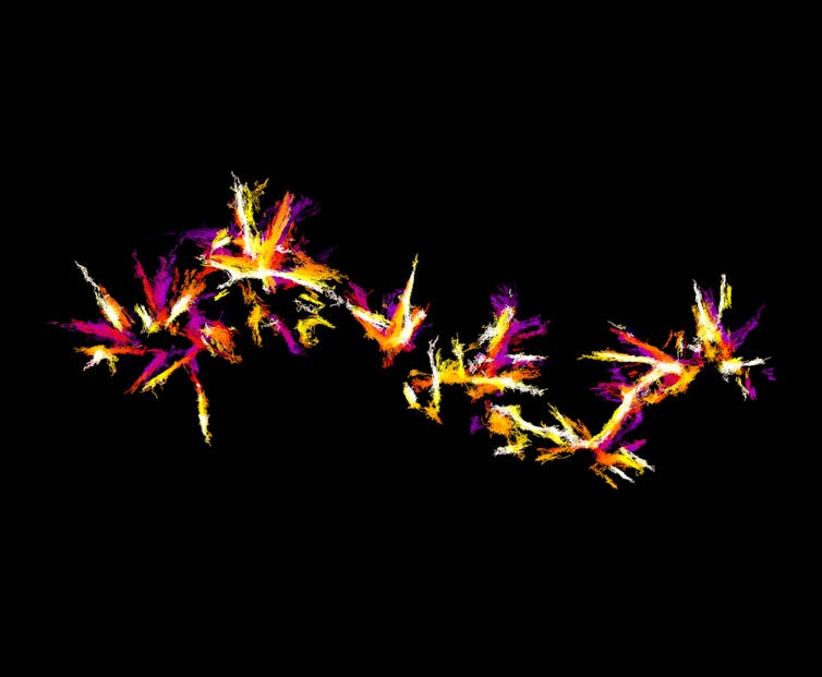
They may look like fireworks, but this image shows nanoscopic movements of single actin molecules. Actin is an essential protein found in all cells of plants and animals, in this case, a neurosecretory cell, a specialised type of nerve cell that releases message molecules into the blood.
Merja Joensuu/QBI
They may look like fireworks, but this image shows nanoscopic movements of single actin molecules. Actin is an essential protein found in all cells of plants and animals, in this case, a neurosecretory cell, a specialised type of nerve cell that releases message molecules into the blood.
 Lee Fletcher/QBI
This image shows the activity of a single neuron (gold) in the brain region the cortex, recorded after the surrounding neurons (cream) are activated with a flash of light.
Lee Fletcher/QBI
This image shows the activity of a single neuron (gold) in the brain region the cortex, recorded after the surrounding neurons (cream) are activated with a flash of light.
 Amandine Grimm/QBI
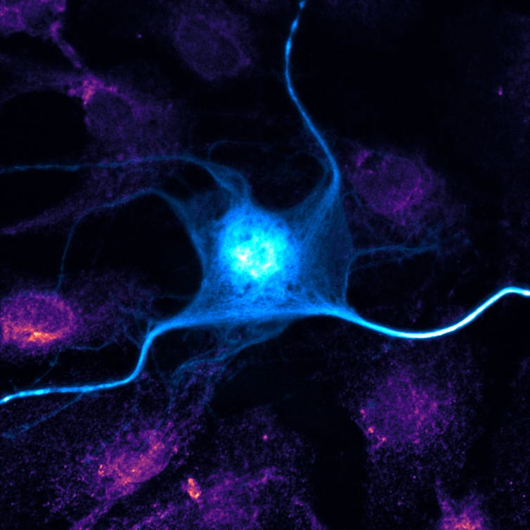
The blue neuron, which could be a manta ray atop a coral reef, expresses a protein tagged with a fluorescent marker. The pink of surrounding cells is formed from endoplasmic reticulum, a cell structure important for processing and transporting proteins.
Amandine Grimm/QBI
The blue neuron, which could be a manta ray atop a coral reef, expresses a protein tagged with a fluorescent marker. The pink of surrounding cells is formed from endoplasmic reticulum, a cell structure important for processing and transporting proteins.
 Eline van de Ven/QBI
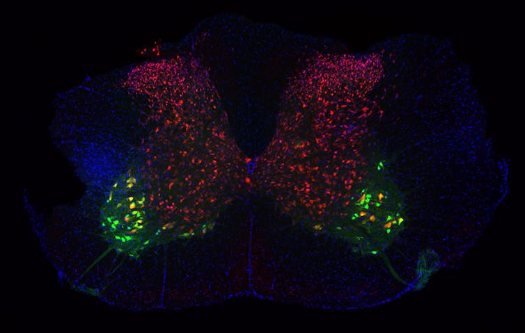
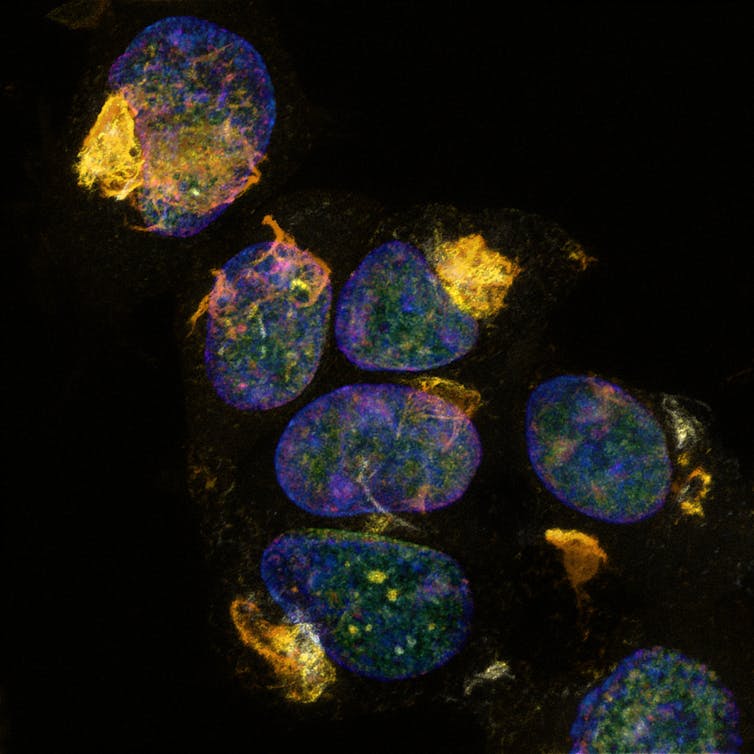
This section of a mouse spinal cord shows a diversity of neuron types. The smaller neurons in pink are involved in pain and the large green neurons are involved in movement.
Eline van de Ven/QBI
This section of a mouse spinal cord shows a diversity of neuron types. The smaller neurons in pink are involved in pain and the large green neurons are involved in movement.
 Amandine Grimm/QBI
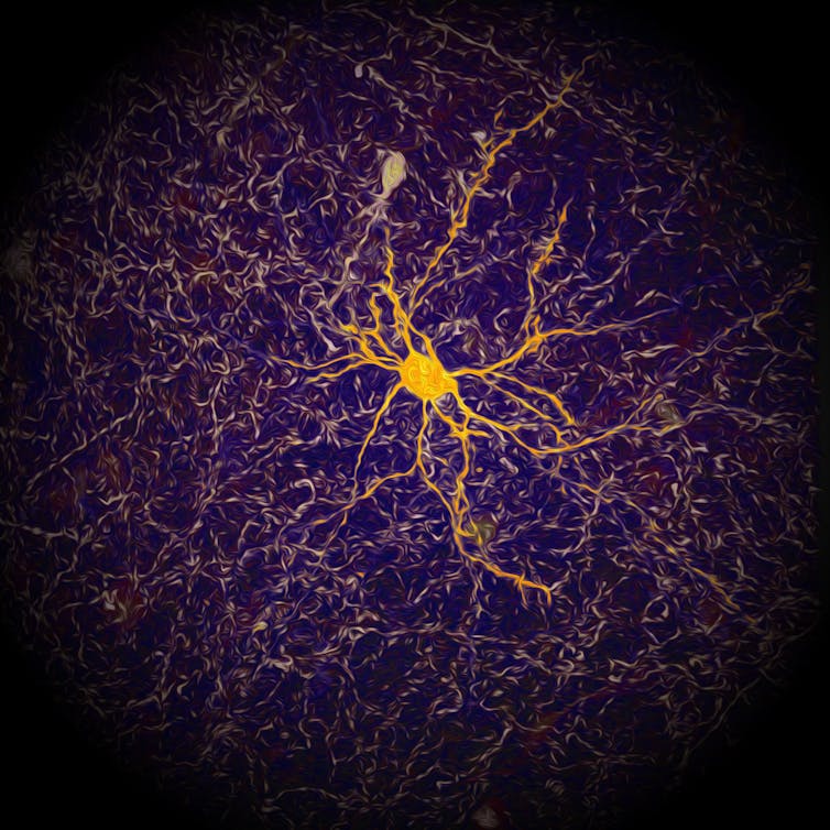
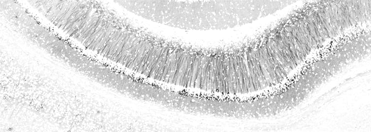
The organisation of neurons in the hippocampus, a brain region important for learning and memory, looks like a forest in snow. The “snow” is made of cell nuclei, which contain each cell’s genetic material. The “trees” are the neurons’ projections, along which electrical signals travel to enable communication with other cells.
The brain in disease
Amandine Grimm/QBI
The organisation of neurons in the hippocampus, a brain region important for learning and memory, looks like a forest in snow. The “snow” is made of cell nuclei, which contain each cell’s genetic material. The “trees” are the neurons’ projections, along which electrical signals travel to enable communication with other cells.
The brain in disease
 Adam Briner/QBI
In Alzheimer’s disease, tau protein (gold) becomes toxic as it builds up. It’s hard to believe these mesmerising, gem-like clusters can be so destructive.
Adam Briner/QBI
In Alzheimer’s disease, tau protein (gold) becomes toxic as it builds up. It’s hard to believe these mesmerising, gem-like clusters can be so destructive.
 Kok-Siong Chen/QBI
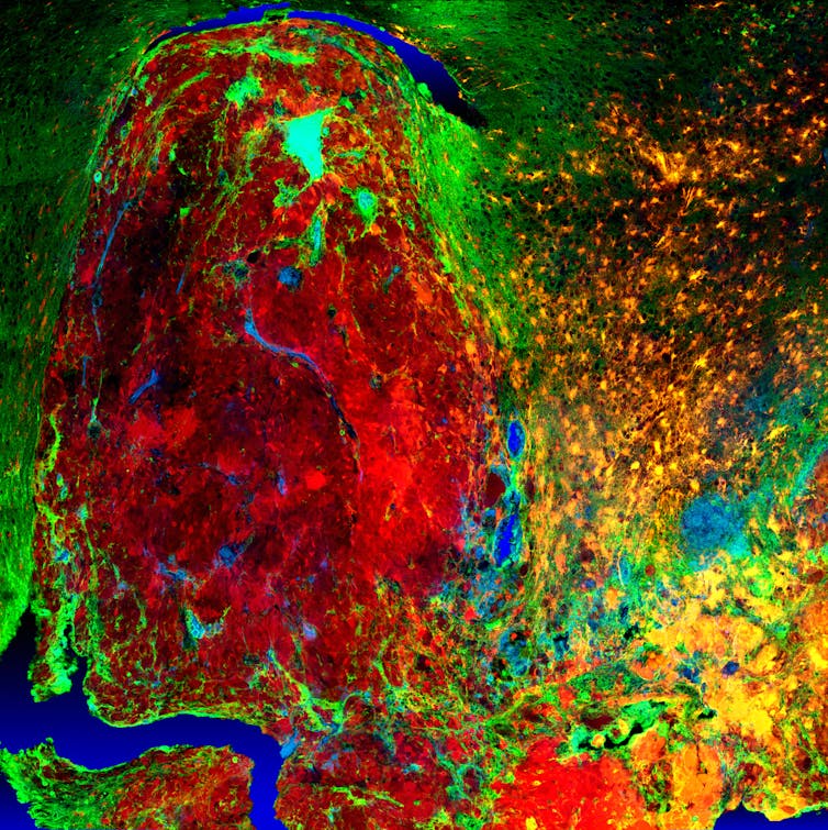
Understanding the characteristics of high-grade brain tumours is crucial to finding treatments for disease. High-resolution fluorescent imaging allows us to investigate how the normal brain cells become cancer cells and how they behave. This image demonstrates the infiltration process of the cancer cells (red) into the normal brain tissue (green).
Insights from nature
Studying model organisms including sea creatures, zebrafish, and roundworms provides insight into vision, brain development, and nerve regeneration respectively.
Kok-Siong Chen/QBI
Understanding the characteristics of high-grade brain tumours is crucial to finding treatments for disease. High-resolution fluorescent imaging allows us to investigate how the normal brain cells become cancer cells and how they behave. This image demonstrates the infiltration process of the cancer cells (red) into the normal brain tissue (green).
Insights from nature
Studying model organisms including sea creatures, zebrafish, and roundworms provides insight into vision, brain development, and nerve regeneration respectively.
 Wen-Sung Chung/QBI
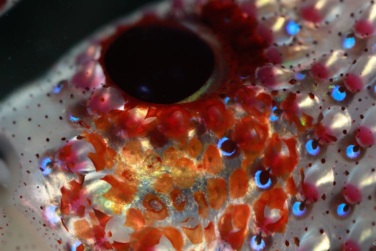
Deep-sea creatures, including this jewel squid, emit their own light for defence, to attract prey, and even to camouflage. At a depth of 600m, the bioluminescent flashes emitted from the light organs of the jewel squid are deadly attractive to prey.
Wen-Sung Chung/QBI
Deep-sea creatures, including this jewel squid, emit their own light for defence, to attract prey, and even to camouflage. At a depth of 600m, the bioluminescent flashes emitted from the light organs of the jewel squid are deadly attractive to prey.
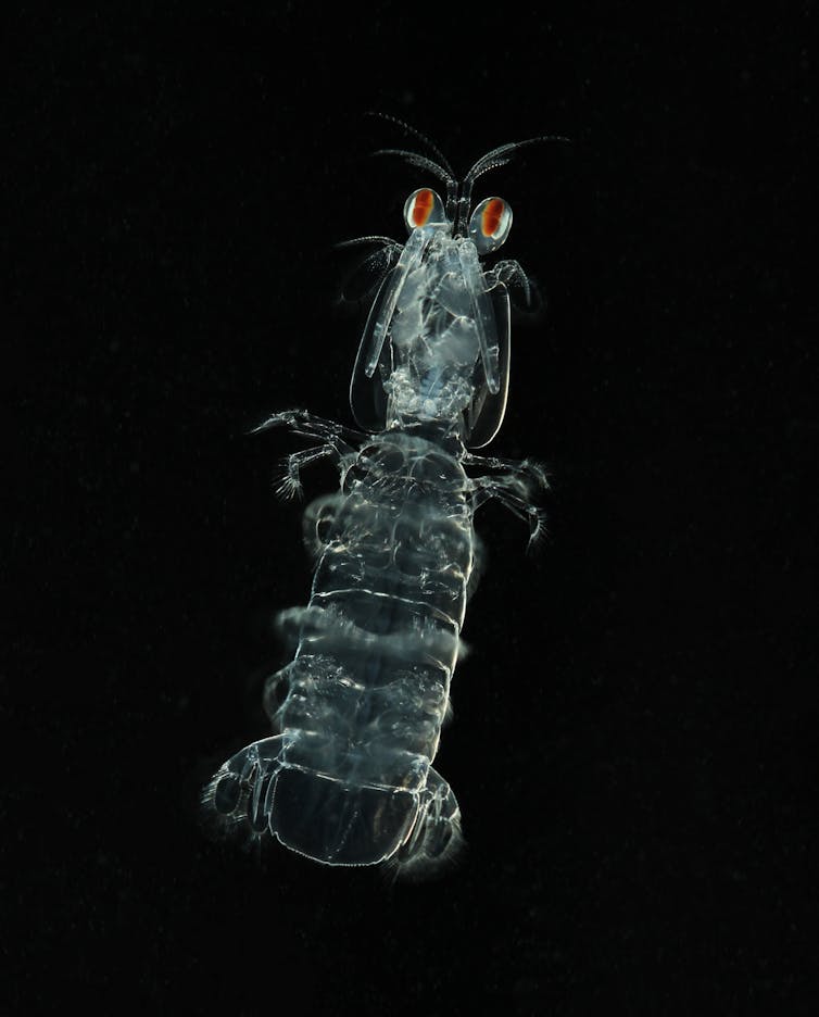 Miriam Henze/QBI
Two retinas are visible in each eye of this mantis shrimp. Mantis shrimp have the most complex visual system in the world; they can see visible and UV light, and can reflect and detect circular polarising light, an extremely rare ability in nature.
Miriam Henze/QBI
Two retinas are visible in each eye of this mantis shrimp. Mantis shrimp have the most complex visual system in the world; they can see visible and UV light, and can reflect and detect circular polarising light, an extremely rare ability in nature.
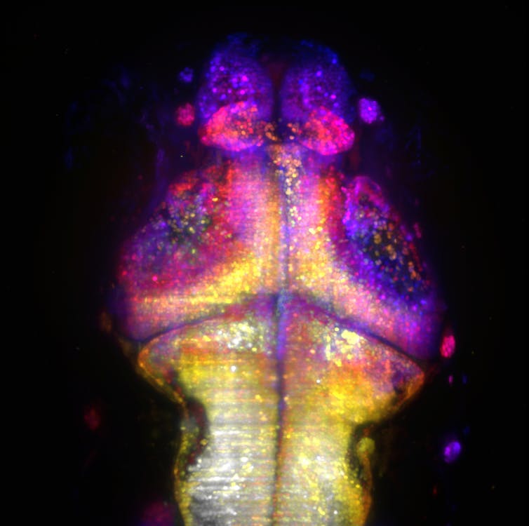 Rumelo Amor/QBI
These are neurons firing in the brain of a one-week old zebrafish, recorded in 3D using a custom-built microscope and colour-coded for depth. Imaging activity in the brains of young zebrafish could lead to an understanding of how the brain is shaped for function.
Rumelo Amor/QBI
These are neurons firing in the brain of a one-week old zebrafish, recorded in 3D using a custom-built microscope and colour-coded for depth. Imaging activity in the brains of young zebrafish could lead to an understanding of how the brain is shaped for function.
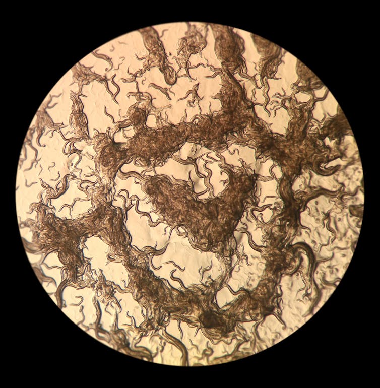 Xue Yan Ho/QBI
A dish of C. elegans roundworms at different stages of their lifecycle. C. elegans is a simple, semi-transparent organism, making it an ideal model for researchers to study the nervous system.
With thanks to QBI graphics designer Dr Nick Valmas, science writer Donna Lu and QBI PhD candidate Abdalla Z Mohamed.
Xue Yan Ho/QBI
A dish of C. elegans roundworms at different stages of their lifecycle. C. elegans is a simple, semi-transparent organism, making it an ideal model for researchers to study the nervous system.
With thanks to QBI graphics designer Dr Nick Valmas, science writer Donna Lu and QBI PhD candidate Abdalla Z Mohamed.
Authors: Wei Luan, Postdoctoral Researcher, The University of Queensland
Read more http://theconversation.com/neuroscience-in-pictures-the-best-images-of-the-year-89077


